Why do eukaryotic cells have organelles?
Compartmentalizing processes allows the cell the maintain different chemical conditions, concentrate certain enzymes and substrates. and prevents incompatible reactions from interfering with one another
| Term | Definition |
|---|---|
Why do eukaryotic cells have organelles? | Compartmentalizing processes allows the cell the maintain different chemical conditions, concentrate certain enzymes and substrates. and prevents incompatible reactions from interfering with one another |
Cytosol | - Site of most metabolic functions and processes
- Site of protein synthesis (translation in eukaryotes and prokaryotes)
- Has a cytoskeleton (network of protein filaments) throughout that provide structural support, maintain cell shape, anchor organelles, facilitate intracellular transport, and enables cell movement. |
Nucleus | - Contains main genome (stores DNA)
- Site of DNA replication
- Site of transcription (DNA -> RNA)
- Main site of ribosome synthesis
- Has a double membrane - outer is continuous with the ER membrane
- Has pores that facilitate the movement of proteins, RNA, etc in and out of the nucleus
- Has heterochromatin (highly condensed chromatin consisting of DNA wrapped around histones) 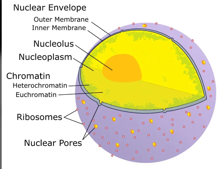 |
Nucleolus | - Non-membrane bound organelle INSIDE the nucleus
- Synthesizes and assembles ribosomes
- Synthesizes rRNA |
Nucleoplasm | Fluid environment inside the nucleus |
Nuclear lamina | Protein network that supports the inner nuclear envelope |
Endoplasmic reticulum | - Main site of lipid synthesis
- Site of synthesis of proteins destined for secretion, membranes, or organelles
- 2 types
Smooth ER - role in lipid synthesis (cholesterol, phospholipids)
Rough ER - Has ribosomes that synthesize proteins |
Golgi Apparatus | - Modifies, sorts and packages proteins and lipids for secretion or delivery to another organelle
- Like a post office
Cis golgi - side that is close to the nucleus
Trans golgi - closer to the cell membrane 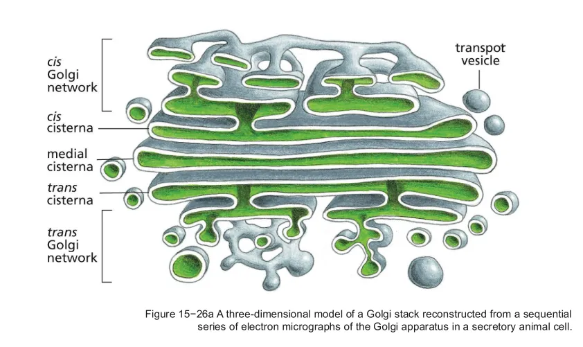 |
Lysosomes | - Site of intracellular degradation (digestion)
- Contains digestive hydrolytic enzymes for breaking down macromolecules, cellular debris, and foreign invaders EX) Nucleases, proteases, glycosidases etc
- Has an acidic internal environment
-Internal hydrolytic enzymes require acidic conditions to be active 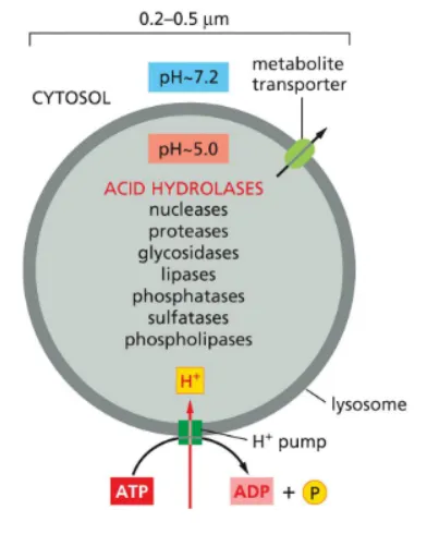 |
Mitochondria | - Site of ATP synthesis by oxidative phosphorylation
- Site of Cellular respiration
- Have own DNA
- Have own ribosomes
- Have double membrane |
Chloroplasts (plants) | - Site of ATP synthesis in plant cells
- Site of carbon fixation in plant cells
- Site of photosynthesis
- Have own DNA
- Have own ribosomes
- Have double membrane |
Endosomes | - Sorts incoming material (endocytosed material)
- Vesicles formed by endocytosis containing endocytosed material fuses with an Endosome so it can sort the material
- Can recycle material (sent to membrane), deliver material to lysosomes (degradation) or are transported across the interior of the cell to another destination (transcytosis) 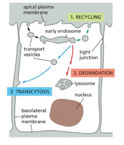 |
Peroxisomes | - Site of oxidative breakdown of toxic molecules
- Can import fully folded and some unfolded proteins (both posttranslational and cotranslational targeting) |
Endomembrane system evolutionary origin | Thought to have arisen through inward folding (invagination) of the ancestral cell's plasma membrane. Related membranes then formed a connected system of compartments and vesicles 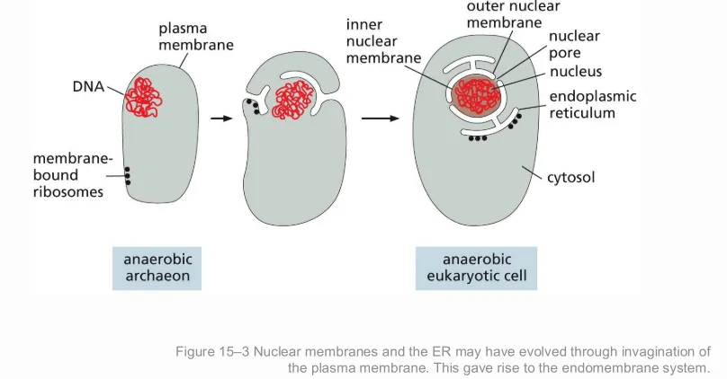 |
Endomembrane system role and components | All components work together to modify, package, and transport lipids and proteins
Nuclear envelope
ER
Golgi apparatus
Endosomes
Lysosomes
Transport vesicles |
Mitochondria and chloroplast evolutionary origin | Endosymbiotic theory - an ancestral anaerobic eukaryotic cell engulfed bacteria that survived inside of it and eventually evolved into organelles
Evidence - Mitochondria and chloroplasts have their own DNA, ribosomes, and divide by binary fission. 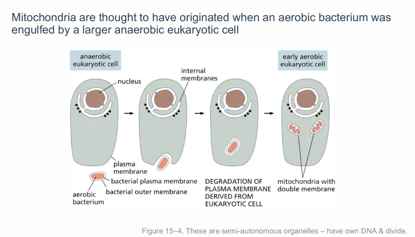 |
3 main ways proteins are transported into organelles and what do all of these processes require? | 1. Through nuclear pores
2. Transport across membranes
3. Transport by vesicles
All require ATP energy |
Targeting signal/sequence | A signal sequence that is a short stretch of amino acids in a protein that direct it to the correct destination in the cell 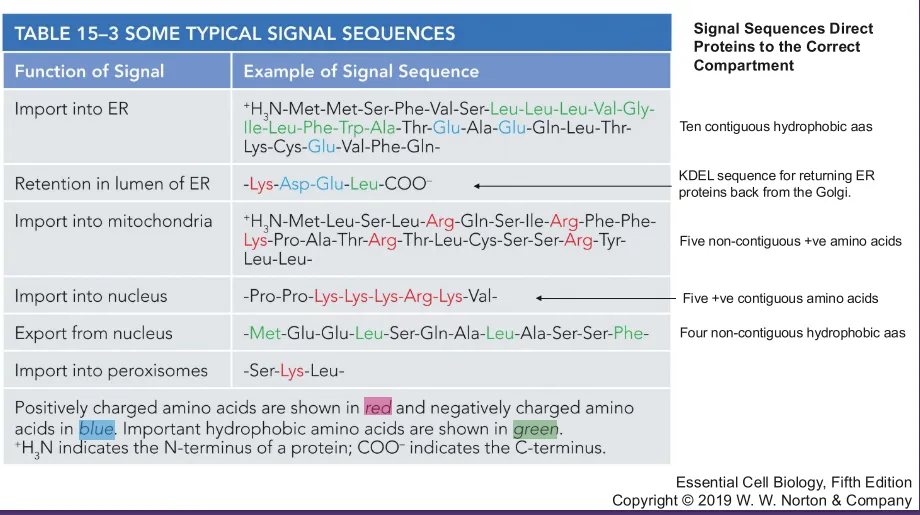 |
2 mechanisms of protein targeting | 1. Post-translational
2. Co-translational |
Posttranslational protein targeting | Targeting that happens after the protein is made
- The protein is fully synthesized, then transported to its destination
EX) Protein transport to the nucleus and transport to mitochondria and chloroplast |
Cotranslational protein targeting | Targeting that happens during translation
- Transport begins while the protein is still being synthesized
EX) ER targeting |
Nuclear pores | Transmembrane protein complexes that act as selective gates located in the nuclear membrane
- Have cytosolic fibrils that select what goes through the pore 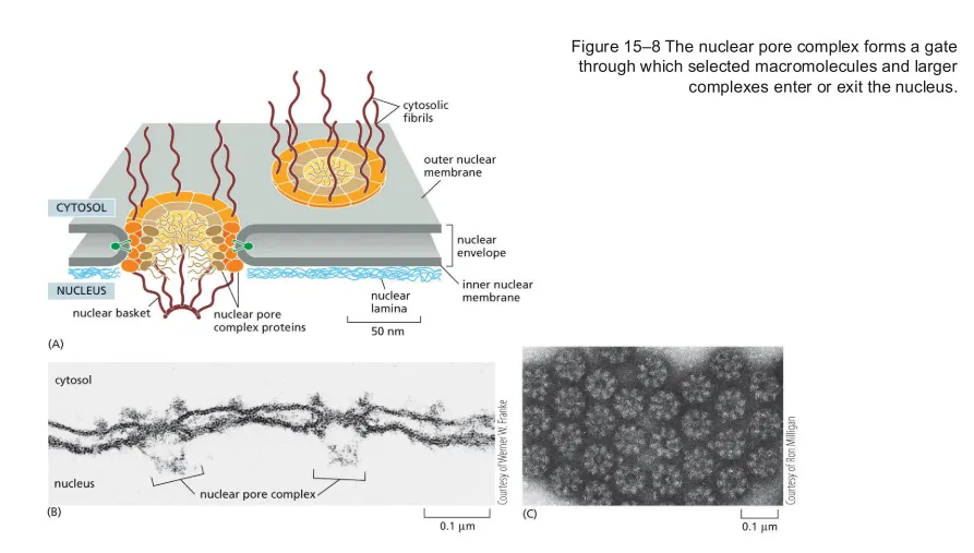 |
Nuclear localization signal (NLS) | A target signal that directs a protein to the nucleus |
Importin | Transport receptor that recognizes NLS on cargo (things that are entering the nucleus)
- Has higher affinity for GTP than cargo it is carrying
- Has weaker interactions with GDP, allowing it to fall off in the cytosol so Importin can pick up more cargo |
Nuclear export signal (NES) | A target signal that directs a protein out of the nucleus |
Exportin | Transporter receptor that recognizes NES on cargo that need to exit the nucleus |
Nuclear transport process | 1. The protein carries an NLS. This signal identifies it as a protein destined for the nucleus.
2. Importin recognizes the NLS and binds the protein
3. The complex interacts with the nuclear pore. The pore recognizes the transport machinery and allows the complex to pass through.
4. Importin dissociates from protein, releasing it to the nucleus, and binds Ran-GTP, then is exported to the cytosol
5. Ran-GAP stimulates hydrolysis of Ran-GTP -> Ran-GDP in the cytosol, causing Ran to dissociate from importin
6. Ran-GDP is transported back into the nucleus and converted back to Ran-GTP by Ran-GEF 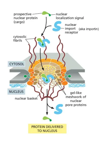 |
GTPase | GTPase is an enzyme that can phosphorylate or dephosphorylate GDP - acts upon GDP
- Ran is a GTPase |
Role of Ran-GTP and Ran-GDP in nuclear import | -Nuclear import, Ran-GTP in the nucleus promotes the release of cargo from importin. The receptor can then return to the cytosol. Importin has a higher affinity for GTP than the cargo it is carrying.
- Ran-GDP in the cytosol promotes its release from importin so importin can be recycled. Ran-GAP stimulates hydrolysis of Ran-GTP -> Ran-GDP in the cytosol, causing Ran to dissociate from importin
Ran protein is a molecular switch that when bound to GTP signals EXPORT (out of nucleus), and when bound to GDP signals IMPORT (into nucleus) 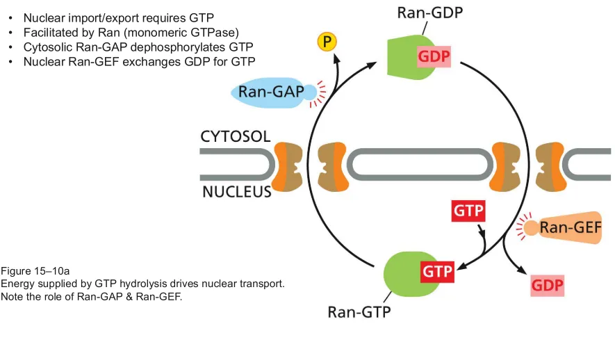 |
Role of Ran-GAP and Ran-GEF in nuclear transport | Ran-GAP - located in the cytosolic side of the nuclear pore and stimulates GTP hydrolysis, converting Ran-GTP to Ran-GDP. - Dephosphorylates GTP
Ran-GEF - located in the nucleus and promotes exchange of GDP for GTP, regenerating GTP
- A high concentration of Ran-GTP in the nucleus and Ran-GDP in the cytosol is maintained to provide directionality to transport 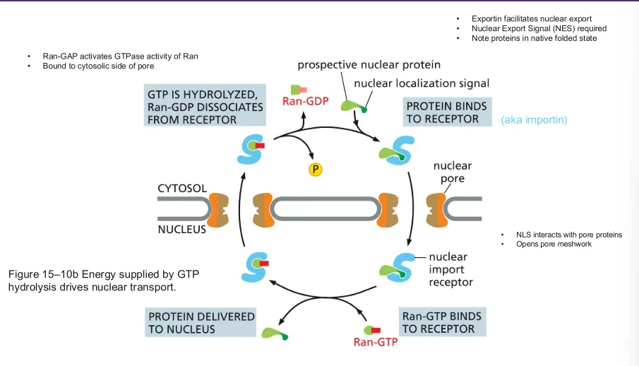 |
In what state is the protein that is crossing the nuclear membrane in? | The protein is in its native, folded conformation |
Name the sub-compartments of the mitochondria and chloroplast 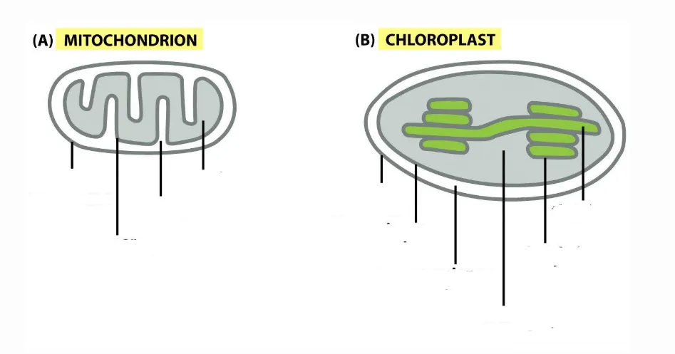 | 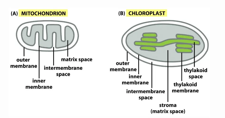 |
Transit signal | Mitochondrial targeting signal sequence on mitochondrial precursor proteins |
TOM and TIM complexes | TOM = translocase of the OUTER membrane - a pore that is trigged once a receptor recognizes the transit signal and transports the protein through the outer membrane
TIM = translocase of the INNER membrane - a pore that helps transport the protein across the INNER membrane towards the matric |
Import into mitochondria process (translocation of proteins into the mitochondria) | *2 membranes must be crossed for a mitochondrial precursor protein to enter the organelle
1. Precursor protein binds to import receptors
2. Receptors trigger TOM complex (pore), protein unfolds as it is ratcheted through TOM, chaperones rachet it through TOM and keep it straight by using ATP
2. Chaperones use ATP to ratchet the unfolded protein through the TIM complex (pore) across the inner membrane towards the matrix
3. Proteins reach their destination in the matrix and fold into functional conformation, and their transit signal is cleaved 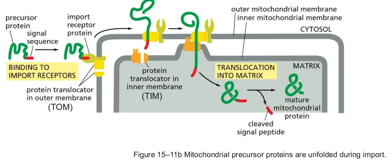 |
Differences between nuclear and mitochondrial import and export | Nuclear
Proteins enter in native folded conformation
Cargo has NLS
Nuclear pores
Mitochondrial
Proteins enter unfolded and refold in the matrix
Chaperones rachet proteins through transport channels
Cargo has transit signal
TOM and TIM complexes |
TOC and TIC | TOC - translocase of the outer chloroplast membrane - helps guide proteins across the outer membrane
TIC - translocase of the inner chloroplast membrane - helps guide proteins across the inner membrane |
Import into chloroplasts process (translocation of proteins into the chloroplast thylakoid space) | * Proteins have to traverse outer membrane, inner membrane, and thylakoid membrane
1. Thylakoid precursor protein carries a signal sequence specific to chloroplast and to thylakoid, chloroplast signal is recognized by membrane receptor - receptor binds protein
2. Receptors trigger TOC complex (pore), protein unfolds as it is ratcheted through TOC by the help of chaperones
3. Chaperones also help the protein to the stoma through TIC using ATP
4. Chloroplast signal is cleaved from the protein once it enters the stoma, allowing for the thylakoid signal to be recognized so the protein can enter the thylakoid 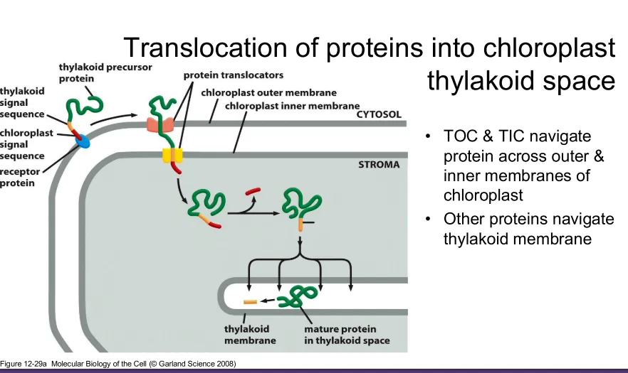 |
In what state is the protein that is crossing the chloroplast and mitochondria membrane in? | Unfolded proteins, one amino acid enters at a time |
2 ways proteins are imported into the peroxisome | 1. From ER budded vesicles
2. Posttranslational translocation from cytosol 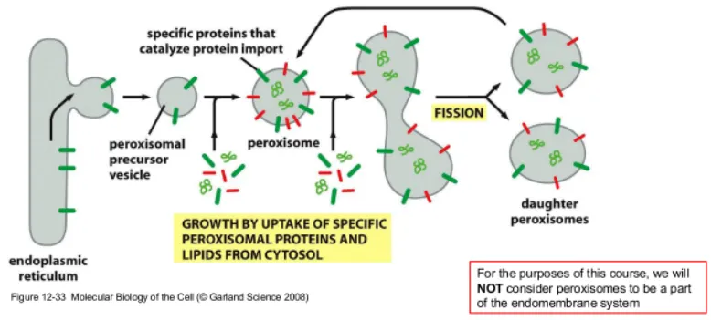 |
In what state is the protein that is crossing the peroxisome membrane in? | Its native, folded conformation |
Is peroxisome protein transport posttranslational or cotranslational | Posttranslational |
Free ribosomes and ER bound ribosomes | Free - float in the cytosol and synthesize proteins that remain in the cytosol or are directed to destinations like the nucleus, mitochondria, chloroplast, and peroxisomes
ER bound - Attach to the ER and translate a protein containing an ER targeting signal. They synthesize many secreted proteins, ER resident proteins, and membrane proteins 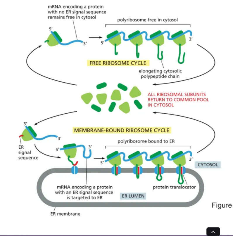 |
Signal recognition particle (SRP) | Molecule made of proteins and RNA that identifies signal sequences on proteins destined for the ER
-Binds signal sequence and the ribosome, pauses translation, and carries the ribosome to the ER to pass it to a SRP receptor
- SRP binding to its receptor happens spontaneously, meaning energy is needed to break them apart so that SRP can be recycled. SRP and its receptor bind GTP, GTP hydrolysis helps release SRP and its receptor after the ribosome is positioned properly |
Cotranslational translocation and posttranslational translocation and their processes | Both describe when a protein crosses the ER membrane relative to its synthesis
Cotranslational - Translation and ER entry happen together
1. Ribosome starts translating mRNA in cytosol
2. Translated ER signal sequence of amino acids emerges and is recognized by SRP, SRP binds the signal and the ribosome, pausing translation
3. SRP brings ribosome to SRP receptor on the ER membrane and positions it over a translocon (protein channel) SRP and its receptor bind GTP, GTP hydrolysis helps release SRP and its receptor after the ribosome is positioned correctly
4. Translation resumes and the N terminus enters first because it was translated first, then as the C terminus elongates, it pushes the rest of the protein through the channel into the ER lumen
- Proteins destined for secretion enter the ER cotranslationally
Postranslational - Translation finishes before ER entry
1. Protein is synthesized in the cytosol
2. After synthesis, protein is directed to the ER using signal sequence (No SRP involved)
3. Protein is transported into the ER through a translocon after translation is complete 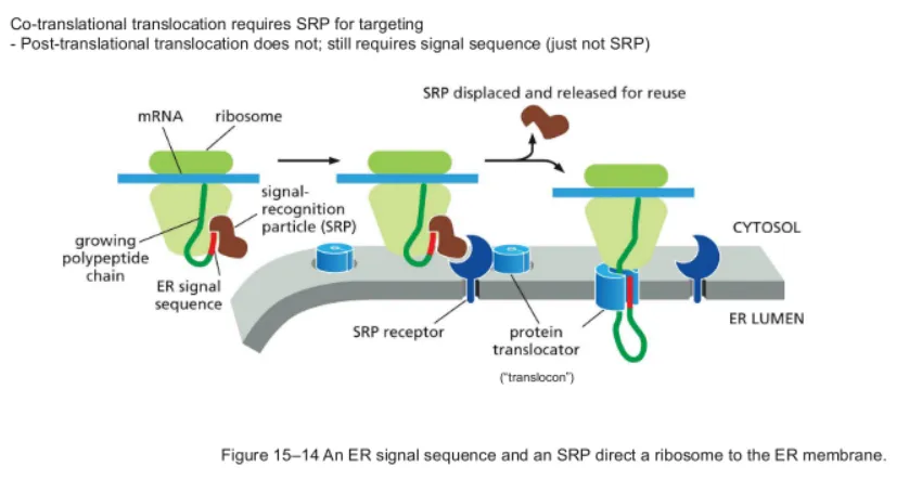 |
How are transmembrane proteins inserted into the ER membrane? | The ER recognizes proteins that should remain in the membrane by start-transfer and stop-transfer signals on the protein
Start transfer signal - initiates the movement of the polypeptide through the translocon
Stop-transfer signal - stops further movement of the segment through the channel and promotes its release sideways into the lipid bilayer |
Single pass transmembrane protein insertion | Internal start-transfer signal initiates translocation, then a later hydrophobic stop-transfer sequence stops passage though the channel and becomes embedded in the membrane. The protein crosses the lipid bilayer ONCE (only stop signal is stuck) 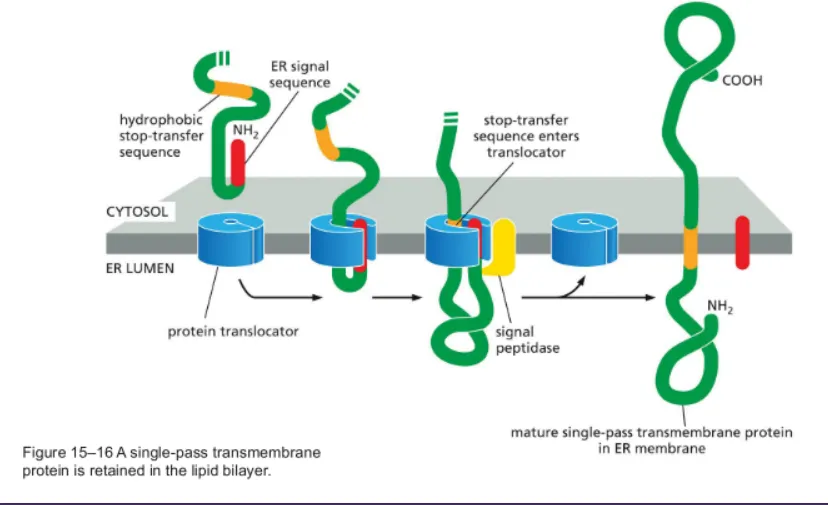 |
Multi-pass transmembrane protein insertion | The double pass protein contains alternating start-transfer and hydrophobic stop-transfer signals, allowing the protein to cross the lipid bilayer multiple times
-Start and stop signals get stuck in the membrane 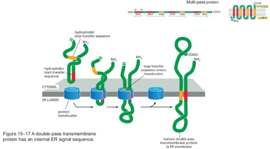 |
Exocytosis and endocytosis (8b) | Exocytosis - Intracellular vesicles fuse with the plasma membrane to release cargo outside of the cell
Endocytosis - Uptake of external substances by engulfing them into vesicles formed from the plasma membrane
- 3 additional types 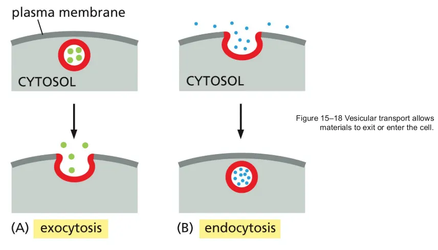 |
What is vesicular transport? | Vesicles are small membrane enclosed sacs that transports substances between compartments of a cell
-Vesicles contain soluble proteins and membrane components 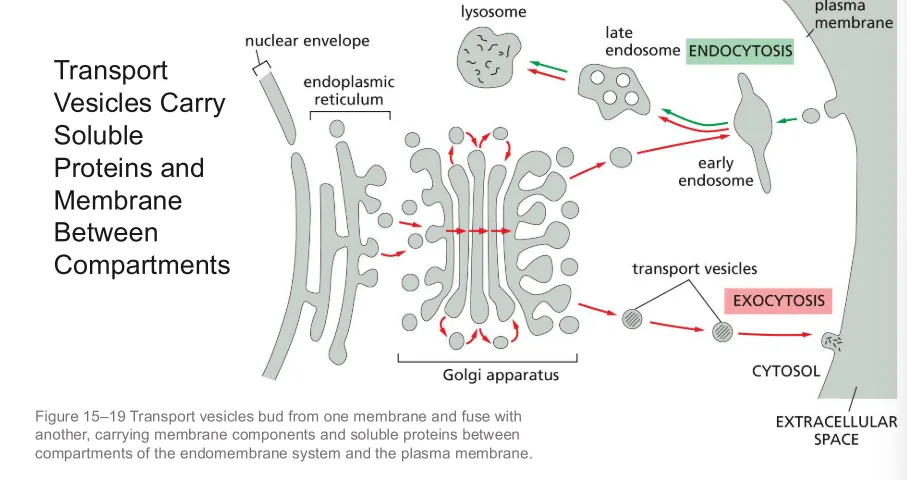 |
Pulse-chase experiment | Technique used to track the movement and fate of a protein inside cells.
- Proteins with a signal sequence are labeled and tracked throughout the cell to see how they go through the endomembrane system 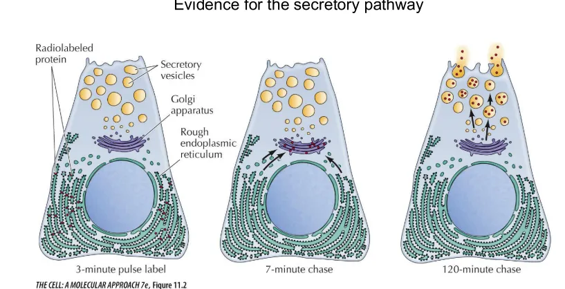 |
Adaptin | Facilitates the interaction between the cargo receptor and the coat proteins |
Protein coats | Proteins that interact with each other to create a mesh coat which allows the vesicle to form
- Helps shape the membrane into a budding vesicle 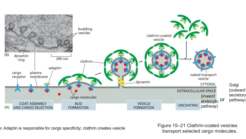 |
How does a vesicle form? | 1. The cell selects proteins and other materials that need to be transported
2. Proteins or materials accumulate at the donor cell membrane and bind to receptors
3. Adaptin is recognized on the cytosolic side of the membrane by the other end of the receptor, allowing coat proteins (clathrins in this example) to assemble on the cytosolic side to help the vesicle overcome the energetically favourable organization of the lipid bilayer so that the vesicle can form around the cargo
4. The vesicle pinches off from the donor membrane (dynamin in this example helps pinch off the vesicle) |
3 groups of proteins that help direct vesicles to their target membranes | 1. Rab proteins (GTP,GDP,GAP)
2. Tethering proteins
3. SNARES (v and t) |
Rab proteins and their 3 forms | Small, monomeric GTP-binding proteins
-Identify the vesicle's destination
-Switch between 2 forms - Rab-GTP and Rab-GDP
Rab-GTP - the active state that can bind a tethering protein
Rab-GDP - the inactive state that releases the tethering protein
GTP is hydrolyzed to GDP, switching off Rab causing it to release its tethering partner so that the machinery can be recycled
Rab-GAP - Activates Rab-GTP to hydrolyze GTP -> Rab-GDP 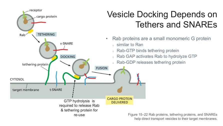 |
Tethering proteins | Proteins located on the target membrane that bind to Rab-GTP on the vesicle and brings it close to the target membrane so that they can fuse |
v-SNARE and t-SNARE | V = vesicle snare (on vesicle)
T = target membrane snare (on target membrane)
- Need ATP to separate them
Proteins that help the vesicle and target membranes fuse by interacting and forming an energetically favourable complex that pulls the two membranes together, thus helping the membranes overcome the energy barrier required to fuse the membranes 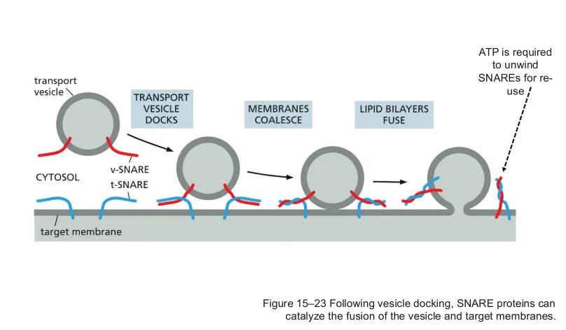 |
Vesicle docking process (3 steps) | 1. Tethering: Rab proteins on the vesicle bind GTP, becoming Rab-GTP, Rab-GTP binds tethering protein on the target membrane to the vesicle, bringing the vesicle close to the membrane
2. Docking: v-SNARE proteins on the vesicle bind to complementary t-SNARE proteins on the target membrane. The proteins pull the two membranes close together. Rab-GAP hydrolyzes Rab-GTP to Rab-GDP, Rab-GDP releases the tethering protein
3. Fusion: v-SNARE and t-SNARE overcome the energy barrier and the membranes merge, creating an opening through which the vesicle contents are released into the target membrane |
Which orientation is the protein in while inside the vesicle during transport? | The cytosolic side of the protein remains the cytosolic side
-Vesicular transport preserves membrane topology
EX) A protein is imbedded in the ER membrane with its N terminus facing the ER lumen and the C terminus facing the cytosol. When it is incorporated into the vesicle, the ER lumen side faces the vesicle lumen, and the cytosolic side remains the cytosolic side |
ER protein modifications | Modifications such as folding and chemical mods are important to protein function
- Can happen cotranslationally or posttranslationally
- Chaperones help proteins fold into the correct shape
- Covalent interactions
*Disulfide bonds = covalent bond between sulfurs
*Glycosylation = covalent attachment of carbohydrate chains to proteins |
Proteasome | Protein complexes responsible for the degradation of proteins into smaller peptides |
What happens to misfolded proteins? | Misfolded proteins are transported to the cytosol, marked for degradation, proteasome recognizes the mark and breaks down the protein 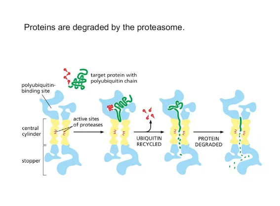 |
What is the UPR? | Unfolded protein response
An accumulation of misfolded proteins in the ER lumen triggers UPR which can:
1. increase chaperone production to increase the cell's capacity to fold proteins correctly (depicted)
2. Reduce the # of proteins entering the ER by decreasing protein synthesis
3. Increase production of mechanisms that remove misfolded proteins (proteasomes) 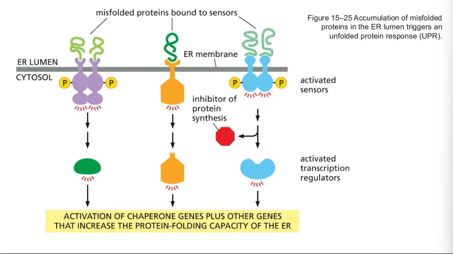 |
Golgi protein modifications | - Addition of oligosaccharides to proteins
- Folding and covalent interactions (similar to ER mods)
|
3 Golgi sorting signals and where they direct the protein | M6P -> Lysosome
GPI -> Apical plasma membrane (facing extracellular space)
KDEL -> ER |
2 main secretory routes from the Golgi | Unregulated exocytosis
Regulated exocytosis
Exocytosis = secretion |
Unregulated exocytosis (secretion) | Vesicles are sent to the membrane
-Vesicles contain membrane components and secretes them without needing a triggering signal
-Helps maintain and renew the plasma membrane |
Regulated exocytosis (secretion) | Cargo in stored in secretory vesicles and are released in response to a specific signal
EX) A hormone-secreting cell may store a hormone in vesicles and release them only when a certain signal arrives |
3 ways to find a protein after it has been labeled | 1. Adding a protease - if the protein was cytosolic it is broken down, but if its in an organelle it is protected. A detergent is then added and the protected protein comes out
2. Centrifugation - if the protein ends up in the sediment its cytosolic, if its in the supernatant its likely in an organelle
3. GFP tagged proteins - GFP produces a fluorescence and is attached to a protein. Transport from the ER to Golgi, to membrane can be tracked (if the tagged protein stops there could be a mutation) |
3 endocytic pathways | 1. Phagocytosis
2. Pinocytosis
3. Receptor mediated endocytosis |
Phagocytosis | Cellular eating - cell reaches out and engulfs large particles like bacteria, dead cells, or debris
- Cell membrane surrounds the particle and forms a large vesicle called a phagosome. The vesicle is then fused with a lysosome |
Pinocytosis | Cellular drinking - cell takes up extracellular fluid
- Membrane forms a vesicle and pinches off into the cell |
Receptor meditated endocytosis | Receptors on the cell membrane bind specific ligands which triggers vesicle formation
- Membrane forms a vesicle and pinches off into the cell |
LDL receptor mediated endocytosis | (Low density lipoprotein) carries cholesterol in the blood
1. LDL binds its receptor on the plasma membrane
2. Receptors and cargo cluster, forming a coated pit with the help of clathrin
3. Vesicle is formed by the pinching off into to cell, bringing LDL and its receptor into the cell
4. Vesicle delivers cargo to endosome which separates LDL from its receptor
5. Cargo and receptors are sorted - receptors are recycled to the membrane and LDL is sent onward for processing and cholesterol release 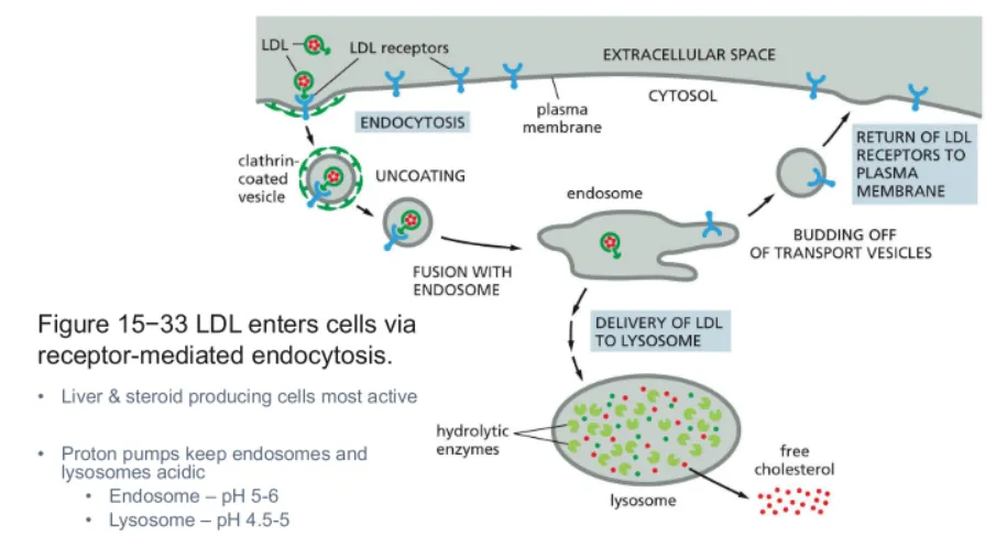 |
3 processes that can happen to materials like receptor proteins after endocytosis (taken up by endosome) | 1. Recycled - materials are returned to proceed with their role like how receptors can be returned to the plasma membrane and can bind more ligand
2. Degradation - materials are sent to the lysosome like how the receptor can be sent to the lysosome to be broken down
3. Transcytosis - materials are taken up and are transported somewhere else |
How is Lysosome's acidic internal environment maintained? | It is regulated by proton pumps
- Protons are pumped into the lysosome, creating a lower pH inside the organelle than the outside
-Internal hydrolytic enzymes require acidic conditions to be active |
Autophagy | Self-eating - Process where components within the cell are marked and sent for degradation
EX) Cell takes its own old, damaged, or unused parts and breaks them down |
3 ways material to be digested reaches the lysosome | 1. Phagocytosis - phagosome vesicle fuses to lysosome and releases its contents
2. Endocytosis - extracellular material is brought in via vesicle and it fuses to the lysosome
3. Autophagy - Old, damaged, or unused components inside the cell are marked for degradation and are engulfed by a autophagosome which fuses with the lysosome
All of these paths require a specific signal |