Structure and function summary
270 bones at birth -> fuse together till 206 as an adult.
Bones are attached to other bones by ligaments.
Bones are attached to muscles by tendons.
Cartilage (articular) covers the ends of bone at a join and allows friction free movement.
| Term | Definition |
|---|---|
Structure and function summary | 270 bones at birth -> fuse together till 206 as an adult.
Bones are attached to other bones by ligaments.
Bones are attached to muscles by tendons.
Cartilage (articular) covers the ends of bone at a join and allows friction free movement. |
Functions of the skeleton | Muscle attachment and movement - via a set of lever, muscles attach to bones and pull on them to create movement.
Storage of minerals - bones act as a rich reservoir for the storage of minerals like calcium and phosphorus to be drawn upon when needed.
Protection of vital organs - the skeleton protects essential organs and delicate structures within the body.
Shape - Gives the body its characteristic shape and framework.
Production - bone marrow cavity of some bones produces blood cells. |
Divisions of the skeleton | Axial skeleton (80 bones) -> trunk of your body
- skull
- rib cage
- spine
- sternum
Appendicular skeleton (126 bones) -> the limbs
- arms
- legs
- pelvis |
Joint classifications - what is a joint? | A joint is where a bone meets another bone.
There are 3 classifications of joint:
Fused/Fibrous - fixed so cannot move (skull, sacrum, coccyx)
Cartilaginous - section of cartilage generally in-between 2 bones (intervertebral discs, symphysis pubis)
Synovial - freely moveable joints (most appendices skeleton, knee, hip, shoulder. |
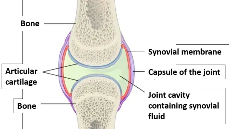
Synovial joints structure | Tendons and ligaments make up the exterior of the joint capsule.
1. Articular cartilage
2. Joint capsule
3. Synovial membrane
4. Synovial fluid
Articular cartilage is used to create smooth movement between bones.
Synovial fluid is what lets things glide.
Cold makes the fluid thicker, warm makes it like olive oil. This is where warming up comes from.
 |
Specific joints and joint actions 01: Glenohummeral joint (shoulder joint) | Humerus bone fits into the scapula bone. The area that they fit together is called the Glenoid fossa. (Very shallow joint, Joint capsule helps to hold ball of the humorous in place.)
Bones to which the muscles attach :
Acroniam (scapula hook on top)
Coricoy process (scapula pointing infront)
Acronym calviular joint -> attaches the scapula and clavicle. Clavicle then attaches to the sternum in a saddle/glide joint.
The stern-cavicular joint, is the only part of which the shoulder girdle attaches to the axial skeleton. 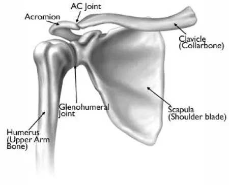 |
Specific joints and joint actions 01: Glenohummeral joint - Glenoid fossa | On the head on the humerus you have the smooth articular cartilage -> also on the surface of the glenoid fossa to make the articulation as smooth as possible.
Shoulder is more easily dislocated due to the shallowness of the glenoid fossa, but also means there is a higher range of movement in the shoulder. 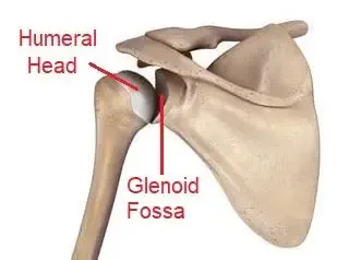 |
Specific joints and joint actions 02/3: Acromioclavicular and Sternoclavicular joints | 2 joints at opposite ends of the clavicle
Sternoclavicular joint : At the sternum end
-> Saddle joint as edge of the clavicle sits in a groove
-> Only connection between the shoulder girdle and the axial skeleton.
Acromioclavicular joint : At the acroniam end (shoulder)
-> articulates the clavicle and acroniam (Scapula top bone). 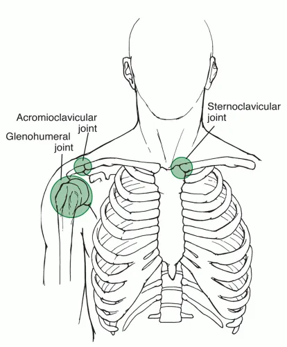 |
Specific joints and joint actions 02/3: Acromioclavicular and Sternoclavicular joints movements | Strenoclavicular joint movements : Saddle/gliding joint
-> Elevation (shoulders go up)
-> Depression (shoulders go down)
-> Protraction (shoulders forward)
-> Retraction (shoulders back)
Acromioclavicular joint movement: Gliding joint
-> Elevation (shoulders up)
-> Depression (shoulders down) |
Specific joints and joint actions 04: Elbow joint | - Top end you have the humerus.
- Condials and epicondials (boney bits at the bottom of the humerus)
- Articular cartilage covering the bone
Humerus articulates with two bones:
-> Radius (lateral aspect)
-> Ulna (Major connection to the elbow joint through a hook (olecranon) which connects to the humerus) When you hit this hook it's called the funny bone.
Actions : Flexion and extension. |
Specific joints and joint actions 05/6: Radioulnar joint (Distal and proximal) | Radius has a circular head as it is a rotational joint.
The joint next to the elbow is the proximal radioulnar joint - closer to the centre of the body.
The joint at the wrist is the distal radioulnar joint - further from the centre of the body.
At proximal the ulna is the more prominent/larger bone but at the distal the radius is the more prominent/larger bone.
Pronation (palm down) and supination (palm up) is the rotational join of the radius and ulna that allows the hand to face up and down.
Gap between ulna and radius is filled with connective tissue (interossas membrane) -> helps with rotational movement. 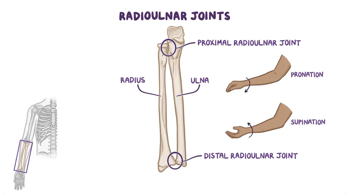 |
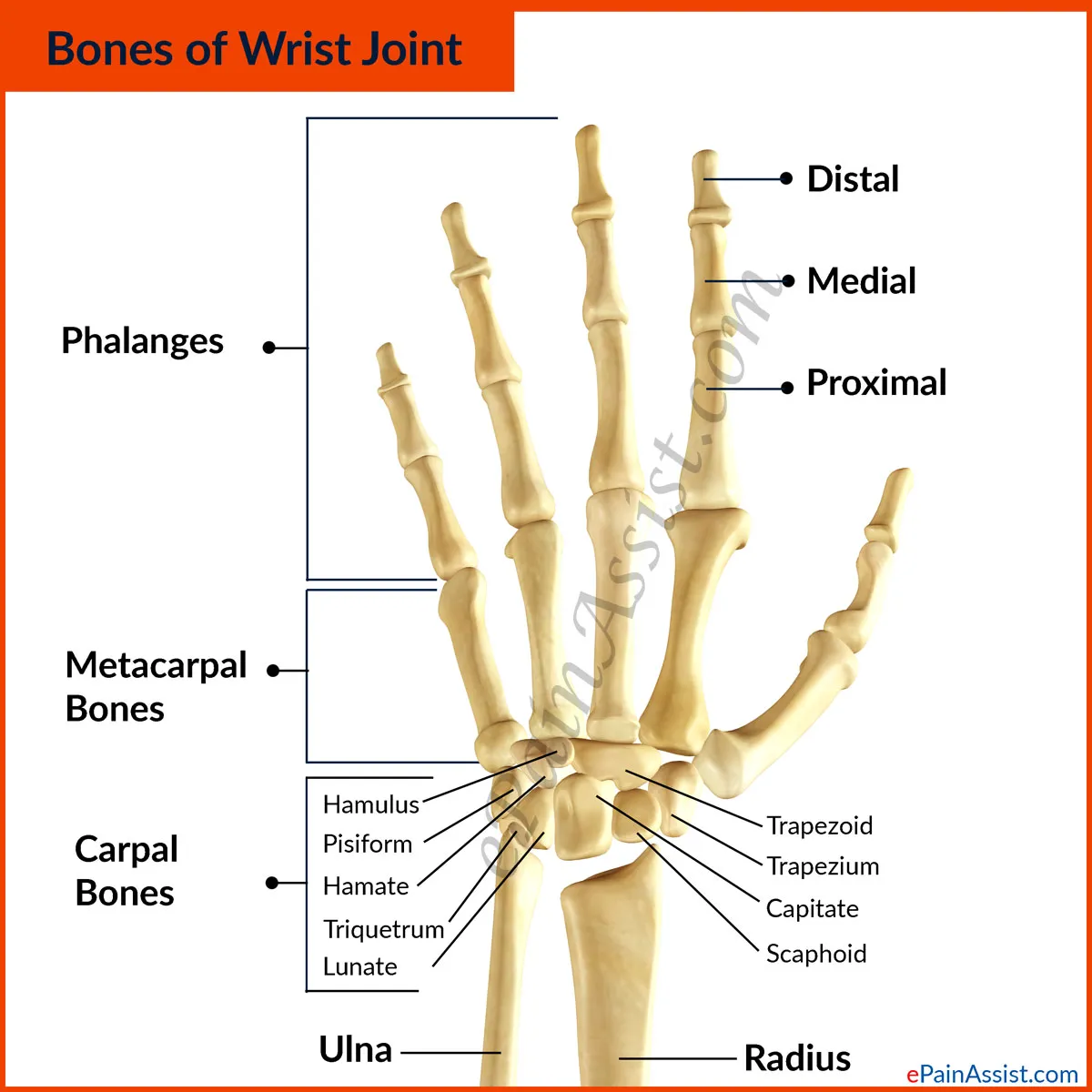
Specific joints and joint actions 07: Wrist Joints | Radial bone has the greater surface area connecting to the wrist (the carpals) -> although the ulna bone sits next to the wrist, it only really articulates with the radius not the carpals.
Carpal bones are gliding synovial joints. As there are a number of them together, meals all the gliding motion gives us the flexion and extension in our wrists. If there was just one joint we wouldn't have the same articulation at the wrist.
Movements at the wrist:
-> flexion (palm to arm)
-> extension
-> abduction and adduction
 |
Specific joints and joint actions 07: Wrist Joints | Saddle joint at the thumb movements:
-> flexion and extension
-> abduction, adduction
->circumduction (not rotation as limited, more of an accumulation of the other movements.) 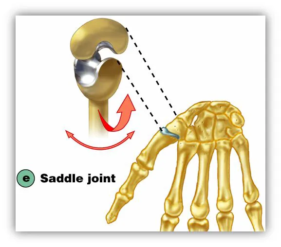 |
Specific joints and joint actions 08: Hip Joint | Head of the femur fits into the side of the pelvis (hip joint).
Pelvis (all fused together creating nominate bone):
-> top of the pelvis is the allium
-> posterior of the pelvis is the ischium (sit bones)
-> front of the pelvis is the pubis
The fusion of these three creates the hip joint/space called the assetabulum, where the head of the femur sits.
It is a lot deeper than the shoulder joint and a lot bigger. Therefore it has better connection, bone on bone. Bigger ligaments, tendons, muscles combine for a stronger joint capsule. Extremely important as all of our weight is on this joint.
Movement (Ball and socket joint)
-> Flexion, extension, adduction, abduction, medially rotate, laterally rotate, circumduction, horizontal extension, horizontal flexion. 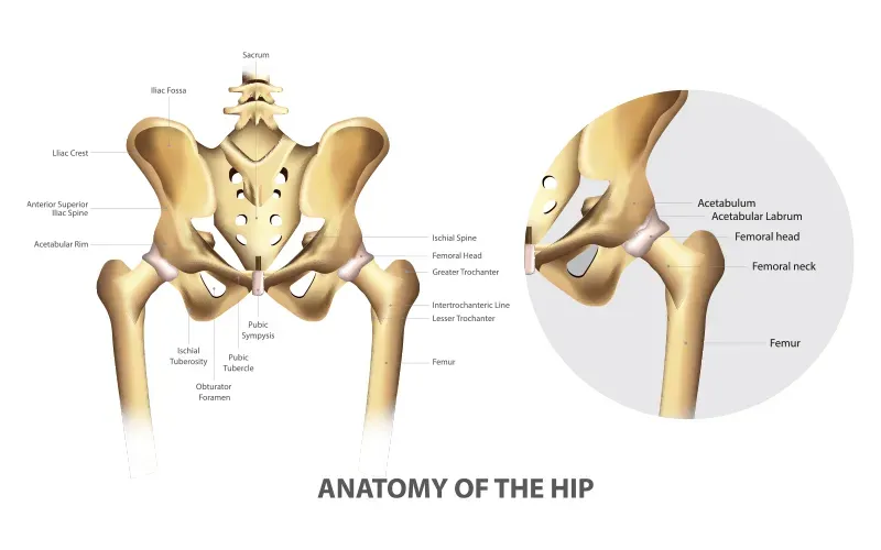 |
Specific joints and joint actions 09: Sacroiliac joint | Joint between the sacrum and the illium.
The surface that the allium sits on the sacrum is called the Ala, which is fairly flat and is quite a thick surface area.
Gliding action, but not too much movement as you want a stable pelvis.
Pregnancy creates a hormone (relaxin), makes lots of the body ligaments relax, including the sacroiliac joint causing pain. When working with pregnant/post-natal have to be weary of this. 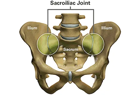 |
Specific joints and joint actions 10: Symphysis pubis joint | Joint between the front aspect of the two parts of the pelvis is the pubic symphysis.
It is a cartilaginous joint, partly moveable. Excessive movement (sports/pregnancy) can lead to a pain syndrome/issue => minimise movement at this joint. 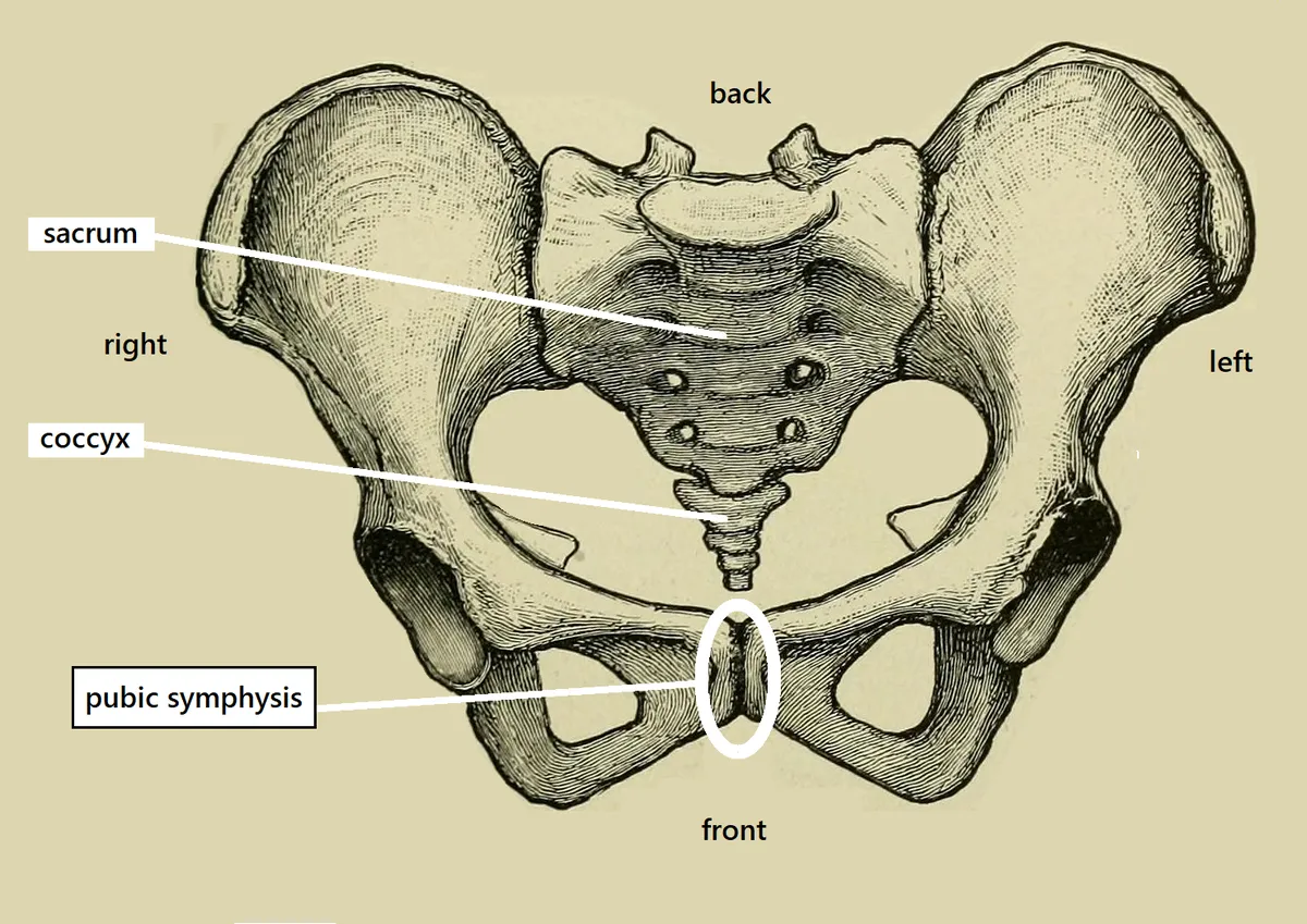 |
Specific joints and joint actions 11: Knee joint | Hinge joint: Flexion, extension and rotation (but only rotate with a professional).
The knee joint is made up of a number of different articulations.
Sesamoid bone -> the patella : only articulates with the femur.
The femur sits on the tibia, with the fibula next to the tibia, however there is no articulation between the fibula and the femur.
Fibula is like scaffolding for the tibia to give it structure. Gap in-between the two bones has connective tissue for stabilisation. |
Specific joints and joint actions 11: Knee joint cont | Articular cartilage of the femur sits on the tibia -> between them there is meniscus cartilage. A c-shaped cartilage that sits on top of the tibia, then the femur sits in that => deeper joint.
Here is where a lot of footballers get injured (meniscus)
Criss cross ligaments in the center of the joint for good stability. 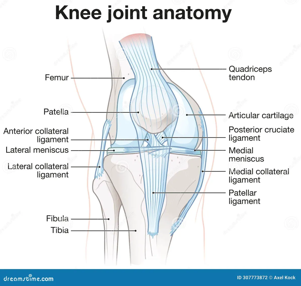 |
Specific joints and joint actions 12: Ankle joint | Fibula comes down and makes the outside of your ankle joint.
Tibia coms down and makes the inside of your ankle joint.
Articulation with the talus (Articular cartilage).
Movement: dorsiflexion (point your toes) plantarflextion (lift toes to the sky).
Talus bone sits on the big heel bone (calcaneus), is your sub-talus joint. Inversion and eversion happens more at the sub-talus joint. 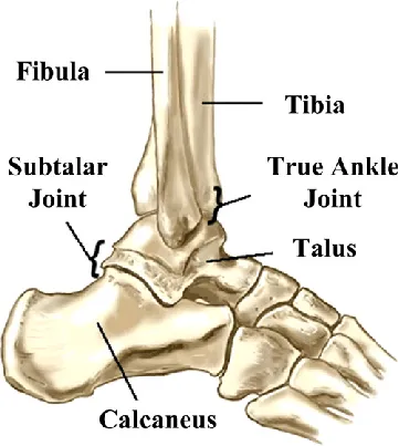 |
Joint action level 3 : Opposition and reposition | Opposition describes the specific movement of touching the thumb to fingers.
Reposition is what makes humans unique from other animals in their ability to grip objects. |