Cortical sensory processing
Created by katie09h8
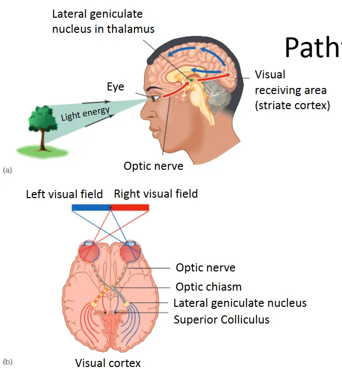
Pathway from Retina to Cortex
Retina → Lateral geniculate nucleus (LGN) → V1
V1: primary visual cortex (occipital lobe), also called striate cortex
From V1: pathways to temporal and parietal lobe → frontal lobe
Here, we focus on processing in (pathway from retina to) V1.
not directly from eye to cortex
early on process is based on visual field
| Term | Definition |
|---|---|
Pathway from Retina to Cortex | Retina → Lateral geniculate nucleus (LGN) → V1
V1: primary visual cortex (occipital lobe), also called striate cortex
From V1: pathways to temporal and parietal lobe → frontal lobe
Here, we focus on processing in (pathway from retina to) V1.
not directly from eye to cortex
early on process is based on visual field
|
Lateral geniculate nucleus processing | LGN cells have center-surround receptive fields. stimulating at sensitive area of the cells, decreases further away
Major function of LGN is to regulate neural signals from retina to
visual cortex.
Signals are received from retina, cortex, brain stem, and thalamus.
Signals are organized by eye, receptor type, and type of environmental information |
Maps: Representing Spatial Layout | Retinotopic map - each place on retina corresponds to a place on LGN
Determining retinotopic maps - record from neurons with an electrode that penetrates the LGN obliquely
Stimulating receptive fields on retina shows location of
corresponding neuron in LGN
gradual shift in position in LGN 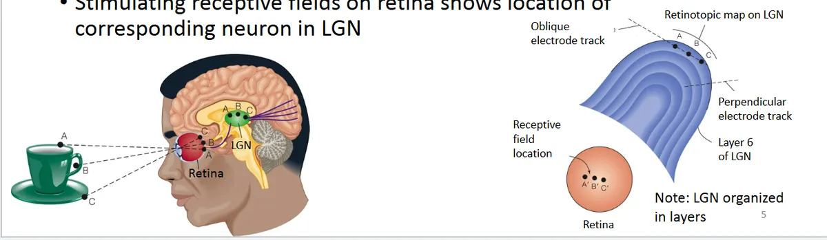 |
The Neural Map on the Striate Cortex (Area V1) | gradually changes in sensitivity to orientation 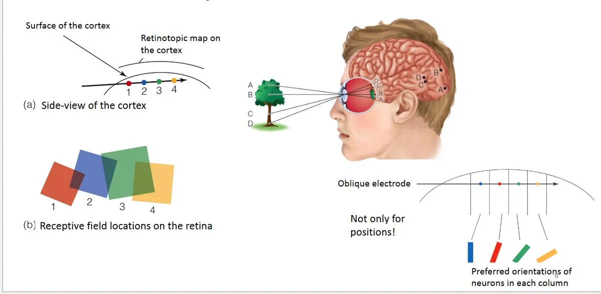 |
Receptive fields | which neurons are active in specific areas of light on retina 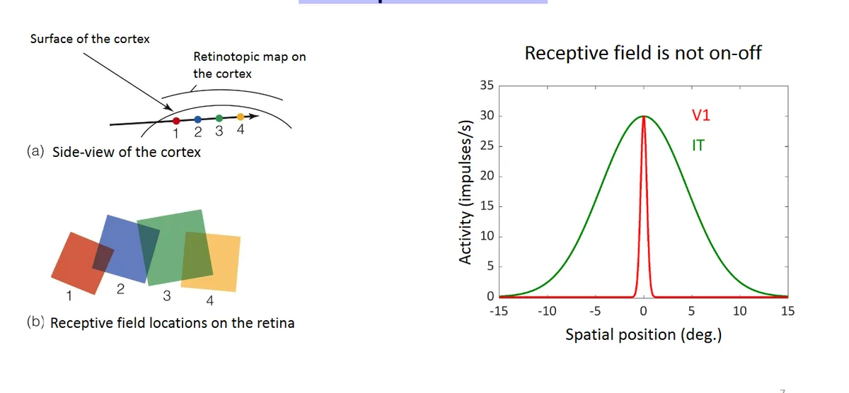 |
fMRI - The Neural Map on the Striate Cortex (Area V1) | Functional magnetic resonance imaging (fMRI)
Hemoglobin carries oxygen and contains a magnetic ferrous molecule
Brain activity takes up oxygen: hemoglobin becomes more magnetic. need blood flow in more active areas, these areas become more magnetic
fMRI: inferring localized brain activity by measuring changes in magnetic response of hemoglobin.
Subtraction technique is used. 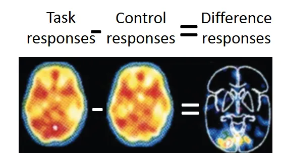 |
Cortical magnification from MRI | Signals from fovea reach larger cortical space (more cells) than
more peripheral parts of retina
more cells processing in fovea, most detailed processing here
where you look you have more acuity 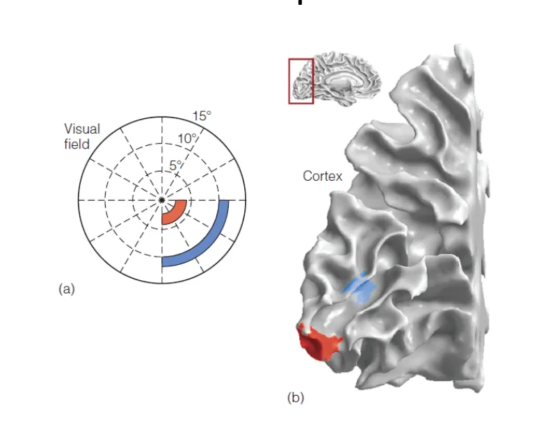 |
Visual Cortex - Location columns | Receptive fields at same location on retina are within a column.
orientation columns are within location columns 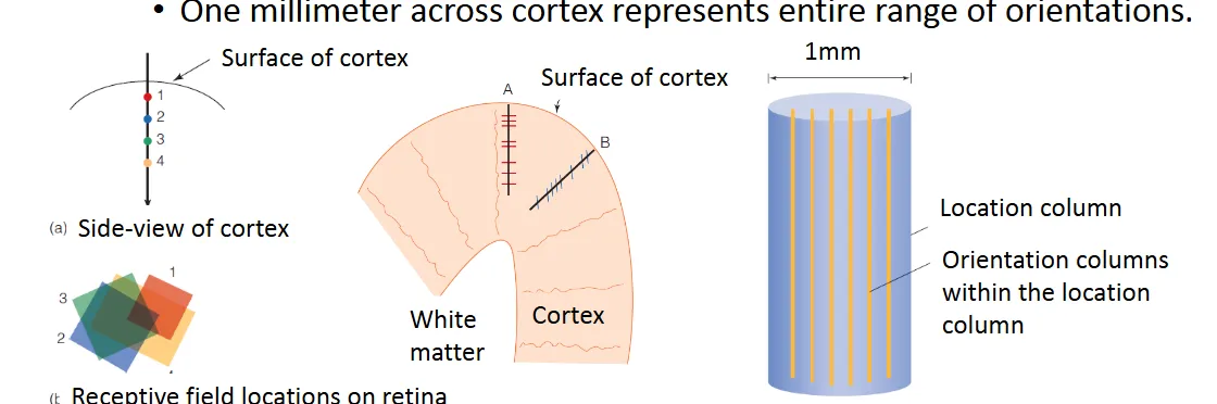 |
Visual Cortex - Orientation columns | Neurons within columns fire maximally to the same orientation of stimuli.
Adjacent columns change preference in an orderly fashion.
One millimeter across cortex represents entire range of orientations.
Spike is an action potential, how active a neuron is
vertical line most active
some cells are tilted, they prefer tilted lines, looks straight to them 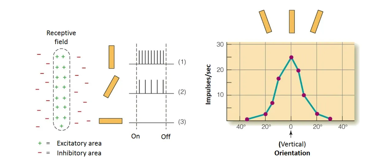 |
Ganglion cell - Receptive Fields of Neurons in the Visual Cortex | Center-surround receptive field. Responds best to small spots but will also respond to other stimuli. |
Lateral Geniculate | Center-surround receptive fields very similar to the receptive field of a ganglion cell. |
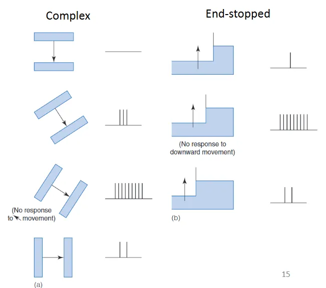
Simple cortical | Excitatory and inhibitory areas arranged side by side. Responds best to bars of a particular orientation |
Complex cortical | Responds best to movement of a correctly oriented bar across the receptive field. Many cells respond best to a particular direction of
movement. |
End-stopped cortical | Responds to corners, angles or bars of a particular length moving in a particular direction
 |
Contextual modulation - “Flexible” Receptive Fields | Perceptual system needs to be flexible.
Kapadia’s (2000) research
Response to stimulation within receptive field affected by ‘what’s happening’ outside receptive field. 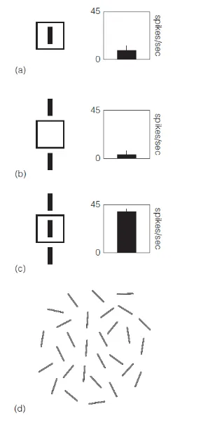 |
Higher-Level Neurons | Inferotemporal (IT) cortex
Prosopagnosia
Fusiform face area
more active when specific complex shapes are present 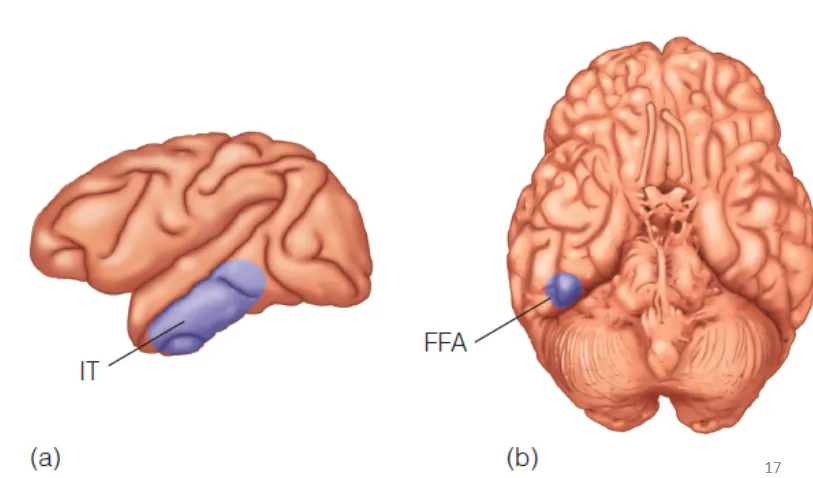 |
Specificity coding | "Grandmother cell” hypothesis
Cells in hippocampus respond to concepts such as Halle Berry.
Problems
Too many different stimuli (people we know, animal names etc) to assign specific neurons
Most neurons respond to several different stimuli
Cell deaths, common theoretically if cell died would lose memory 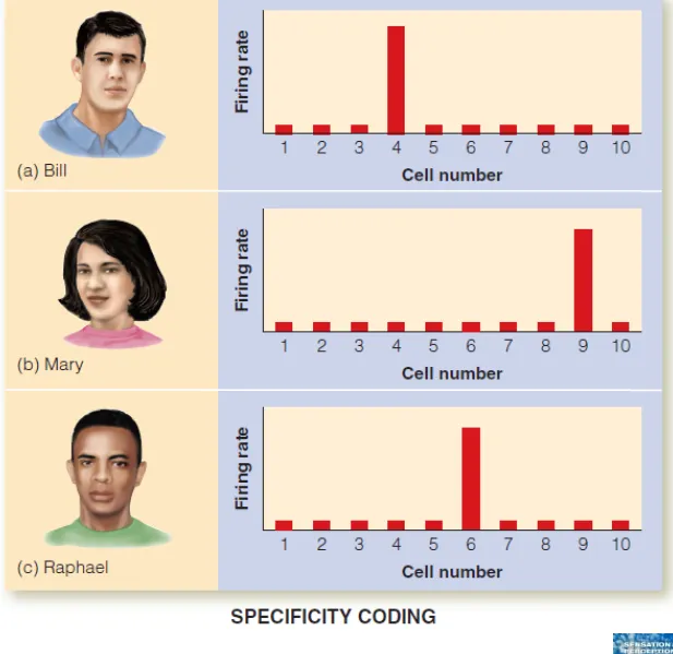 |
Population coding | Pattern of firing across many neurons codes specific objects
Large number of stimuli can be coded by a few neurons. 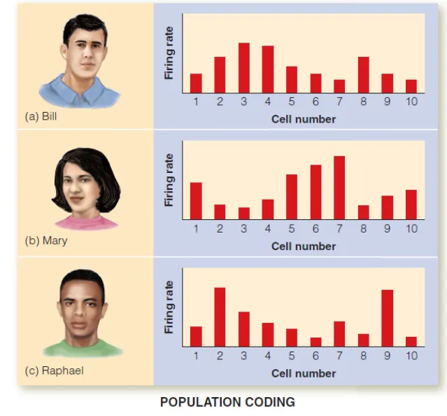 |
Sparse Coding | How many neurons are needed for an object in distributed coding?
Sparse coding: only a relatively small number of neurons are necessary
This theory can be viewed as a midpoint between specificity and distributed coding. 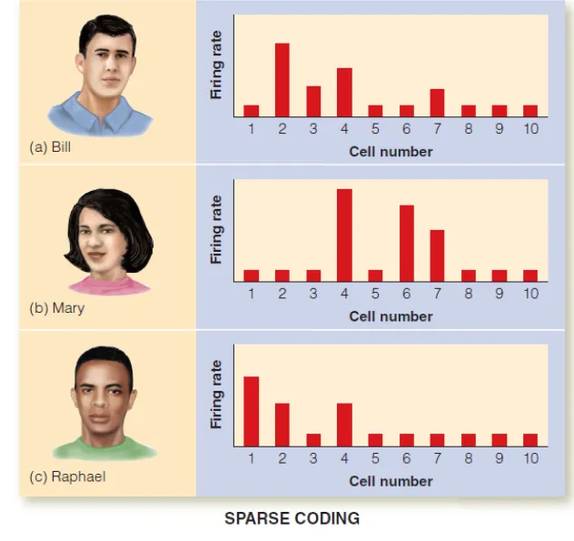 |
Modularity | Module: brain structure that processes information about specific stimuli
Rolls measured response neurons in inferotemporal (IT) cortex in monkeys
Responds best to faces with little response to non- face stimuli
Temporal lobe damage in humans results in prosopagnosia.
Rolls & Tovee (1995)
if this area of the brain becomes damaged won't recognise faces 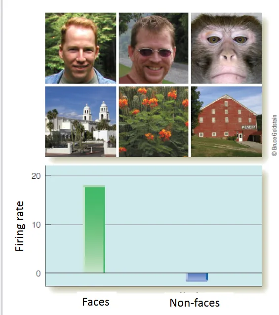 |
Areas for Places, and Bodies in Human Brain | Evidence from humans using fMRI and the subtraction
technique show that:
Parahippocampal place area (PPA) responds most to spatial layout
Extrastriate body area (EBA) responds most to pictures of bodies
and body parts |
selective Adaptation | Neurons tuned to specific stimuli fatigue when exposure is long.
• Fatigue or adaptation to stimulus causes
• Neural firing rate to decrease
• Neuron to fire less when stimulus immediately presented again
• ‘Selective’ means that only those neurons that respond to specific stimulus adapt.
Measure sensitivity to range of stimulus with one same characteristic
Adapt to that characteristic by extended exposure
Re-measure the sensitivity to range of stimulus characteristic
|
Gratings selective adaptation | Alternating light and dark bars
Angle relative to vertical can be changed to test for sensitivity to
orientation.
Difference in intensity can be changed to test for sensitivity to
contrast
Measure contrast threshold by decreasing intensity of grating until person can just see it.
Calculate the contrast sensitivity by taking 1/threshold.
If threshold is low, person has high contrast sensitivity. 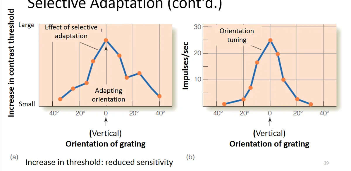 |
Selective Rearing | Animals are reared in environments that contain only certain types of stimuli.
Neurons that respond to these stimuli will become more predominate due to neural plasticity.
Blakemore and Cooper (1970) showed this by rearing kittens in tubes with either horizontal or vertical lines.
Both behavioral and neural responses showed development of predominant sensitivity to these environmental stimuli. 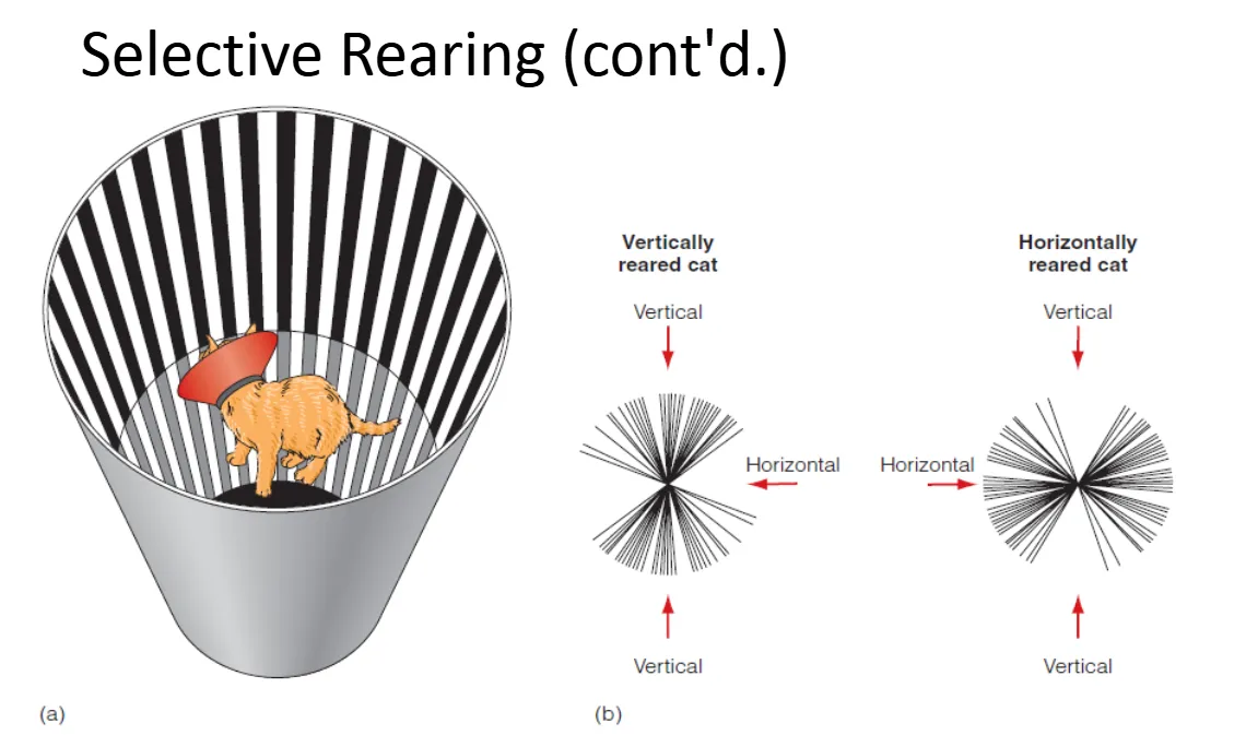 |